PRISM Oncology Series · RWID Multimodal
Breast Screening Images & Outcomes
Breast screening cohort built around digital breast tomosynthesis (DBT), with biopsy-confirmed outcomes, prior and follow-up breast imaging, and free-text radiology reports from routine clinical practice.
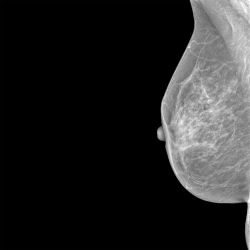
Reconstructed slice from a digital breast tomosynthesis (DBT) screening exam. De-identified.
558
Patients in cohort
271
Cancer cases
287
Non-cancer cases
2017-24
Time frame coverage
Dataset Specifications
What is in the PRISM Breast Screening Images & Outcomes Dataset
Screening and diagnostic breast imaging linked to biopsy-confirmed pathology, receptor status, and free-text radiology reports, with prior and follow-up exams aligned to the index DBT study.

Therapeutic Area
Oncology / Women's Health
Disease Focus
Breast cancer · screening and diagnostic population · BI-RADS 0, 1, and 2 · biopsy-confirmed malignant and benign cases included
Cohort Composition
271 cancer cases, all with pathology results; and 287 non-cancer cases
Imaging Modalities
Digital breast tomosynthesis (DBT) · mammography · breast MRI · breast ultrasound
Clinical Data (EHR)
Demographics · free-text radiology reports · pathology and IHC reports · receptor status (ER, PR, HER2)
Data Sources
Radiology (DICOM) · pathology · nuclear medicine (DICOM)
Data Format & Compliance
Raw DICOMs and structured clinical data · HIPAA de-identified · Provenance documentation built into acquisition · Prior and follow-up breast exams linked to the index DBT study
Cohort Composition
Four outcome groups across the screening population
Every case is stratified by biopsy-confirmed outcome. The malignant and benign balance supports training, validation, and performance benchmarking without additional cohort assembly.
Cancer cases
271 patients: malignancy confirmed on biopsy, with linked pathology reports, IHC, and receptor status (ER, PR, HER2).
Non-cancer cases
287 patients: non-cancer outcome confirmed on biopsy or through resolved screening callback, with linked imaging, radiology reports, and pathology.
Applications
What this dataset enables
From AI model development to screening performance analysis and radiogenomic research.
AI model development & validation
Train, enrich, or validate models for lesion detection and characterization on DBT, using biopsy-confirmed ground truth across malignant and benign cases.
DBT screening performance
Characterize real-world tomosynthesis performance against biopsy outcomes, including recall rates, BI-RADS distribution, and detection across breast density categories.
Missed cancer analysis
Review prior exams against subsequent malignant findings to characterize interval and missed cancers, supported by linked prior and follow-up breast imaging.
Post-screening risk modelling
Build risk models for subsequent malignancy following a negative or indeterminate screening exam, using longitudinal imaging and confirmed outcomes.
Radiogenomic & radiomics research
Link quantitative imaging features to receptor status (ER, PR, HER2) and pathology for radiomic signature discovery and imaging–molecular correlation.
Patient stratification & clinical development
Stratify screening populations by imaging phenotype and receptor status to support clinical development and post-marketing surveillance in real-world settings.
Biomarkers & Endpoints
Key biomarkers and endpoints available for selection
Imaging, pathology, and molecular variables with linked prior and follow-up exams. Variable selection is tailored to your programme's endpoints and research objectives.
Breast Imaging
- BI-RADS score (0, 1, 2)
- DBT lesion characteristics
- Lesion location and size
- Breast density
- Prior and follow-up exam comparison
Pathology
- Biopsy outcome (malignant / benign)
- Pathology reports
- IHC reports
- Histologic subtype
Molecular Markers
- Estrogen receptor (ER)
- Progesterone receptor (PR)
- HER2
Reports
- Free-text radiology reports
- Screening versus diagnostic context
- Recall and callback status
Dataset Request
Request access to PRISM Series RWID Multimodal Cohorts
Receive technical specifications for the pre-built research cohort relevant to your programme.


