


Cancer Centers Bring Multi-Dimensional Support to Real-World Data and Healthcare Research
Reading time /
3 min
Company News
TL;DR
- Cancer centers generate huge volumes of imaging data through routine patient care. Historically, little of that imaging data reached the researchers and innovators building the next generation of diagnostic and therapeutic tools.
- Healthcare institutions, including cancers centers, support real-world data research in different ways: contributing imaging data, or investing in the companies and platforms that streamline this work. Fox Chase Cancer Center, and its parent, Temple Health, supports ground-breaking innovation through its partnership with Segmed, providing both data and investment to improve health outcomes.
Every mammogram, PET scan and MRI ordered at a cancer center adds to an archive that already holds years of imaging history. Most of it serves one patient, once, then sits unused. Real-world data partnerships are built to change that, whether an institution contributes data directly or financially backs the platforms making the infrastructure possible. Fox Chase Cancer Center is an example of an organization who wants to advance innovation by both sharing its rich data (after being de-identified) and, through its investment in Segmed (read the Series A announcement here, from September 2024).
The Imaging Archive Every Cancer Center Already Has
Imaging is central to oncology. It is used for screening, diagnosis, staging, treatment planning and follow-up, a single patient's course of care can produce years of longitudinal imaging, on its own, this archive mostly serves the patient it came from. It rarely reaches the researchers and AI developers working on the next generation of diagnostic tools, especially not at scale.
How De-Identification Makes Data Sharing Possible
Real-world imaging data partnerships exist to close that gap, without exposing the identity of a single patient in the process.
Before any imaging data leaves an institution, it goes through de-identification. Segmed's Incognito tool accurately redacts protected health information from DICOM imaging files, the meta data, and text-based medical reports. The process meets both HIPAA Safe Harbor and Expert Determination standards, the frameworks regulators use to define de-identified health data. De-identification is the key step that lets an institution's imaging archive support outside research, while every patient behind that archive stays unidentifiable.
Fox Chase Cancer Center's Role
Fox Chase Cancer Center was founded in 1904 as the American Oncologic Hospital, the first hospital in the country dedicated to cancer care. It has held National Cancer Institute Comprehensive Cancer Center designation since 1974. In September 2024, after agreeing to provide its imaging data to Segmed, Fox Chase also joined Segmed's Series A round as an investor, alongside a group of healthcare and technology investors.
That investment and high quality data supports Segmed's mission to expand the utility of imaging data which is used to engineer today’s medical innovations. Fox Chase is leading the way and helping improve outcomes for patients more broadly.
“At Fox Chase, accelerating discovery to delivery means finding new ways to put our knowledge and resources to work for patients. Our investment in Segmed reflects that commitment. By responsibly unlocking the research potential of medical imaging data, we can help accelerate innovation in cancer detection, diagnosis, and treatment, benefiting patients at Fox Chase and beyond our walls.” — Robert G. Uzzo, MD, MBA, FACS, President and CEO, Fox Chase Cancer Center-Temple Health
From Imaging Archive to Research & Innovation in Cancer Care
Real-world data from Segmed's network supports innovation along the full regulatory path, from AI algorithms seeking FDA clearance to new therapies advancing toward clinical trials. More than 50 FDA-cleared AI models now used in clinical care were built with Segmed network data, and the same data is helping therapeutics researchers generate the evidence needed to bring new approaches into clinical use.
Oncology is one of the most active areas for this work. Imaging linked to pathology, treatment and outcome data lets developers and researchers follow a patient's course from first scan to confirmed result, and build the evidence regulators look for, whether for a diagnostic tool or a treatment decision aid. Three current cancer programs show how that plays out, from screening to treatment.
Smarter Risk Assessment for Inconclusive Mammograms
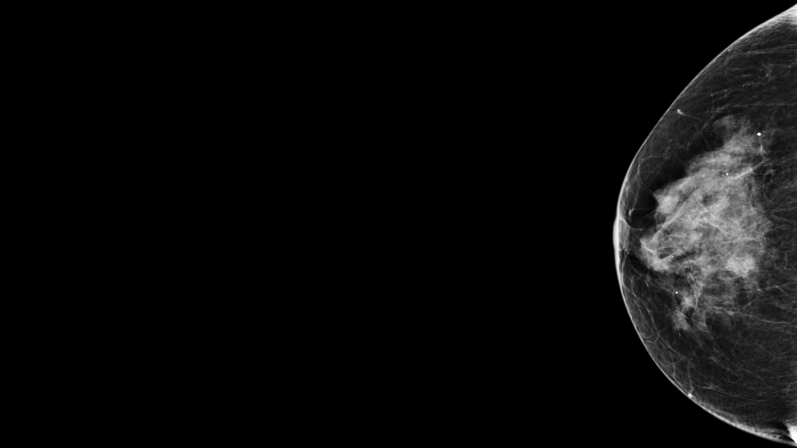
Some breast cancers go undetected in routine screening, and many mammograms come back inconclusive. Using screening and diagnostic mammograms linked to pathology reports from Segmed, an AI developer is clinically validating an algorithm that flags women at higher risk of cancer.
The goal: Earlier detection and fewer unnecessary callbacks.
Real-World Data to Predict Treatment Response in Colorectal and Kidney Cancer
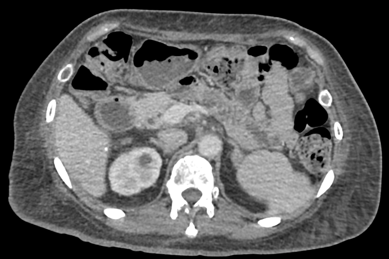
For patients with metastatic colorectal cancer, it can be hard to know early on whether a treatment is working, and real-world scans rarely cover the same parts of the body. Using serial CT imaging linked to outcome data from Segmed, researchers evaluated a deep learning biomarker that predicts overall survival from baseline and early on-treatment scans, even when anatomic coverage varies. The same biomarker was also tested in patients with metastatic kidney cancer treated with immunotherapy, where it showed an even stronger prognostic signal. The results were presented at American Society of Clinical Oncology (ASCO) Meeting 2026. Read the abstract.
The goal: Earlier insight into whether a treatment is working.
From Screening Scan to Confirmed Diagnosis: Training AI for Earlier Lung Cancer Detection
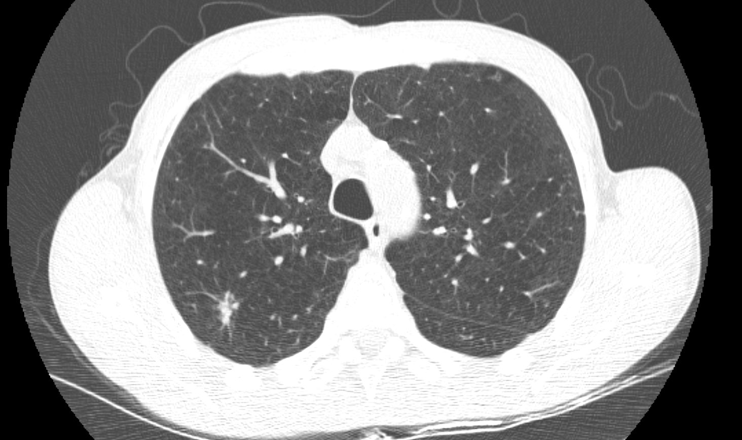
Lung cancer screening finds many nodules. Most are benign, and deciding which ones need action is hard. Using screening CT exams linked to follow-up imaging and pathology reports from Segmed, an AI developer is building an algorithm to detect lung cancer earlier.
The goal: Faster diagnosis and fewer unnecessary procedures.
For institutions that contribute data directly, an unused imaging archive becomes a resource for outside research, with de-identification protecting patient privacy throughout. Institutions can support the same goal also by backing companies and platforms financially.
Segmed builds the infrastructure that turns imaging archives into a resource for outside research. Institutions and investors, including Fox Chase Cancer Center, help expand what that infrastructure can do. Learn how Segmed collaborates with health providers to activate their imaging data. Schedule a call with us.
Frequently Asked Questions - F.A.Q.
What is real-world imaging data?
Real-world imaging data is medical imaging, like MRIs, CT scans, and mammograms, collected during routine patient care rather than inside a clinical trial. It reflects how disease and treatment look across a broad patient population.
How is patient privacy protected in imaging data research?
Imaging and reports are de-identified before they leave the healthcare institution. Tools like Segmed's Incognito remove protected health information following HIPAA Safe Harbor standards.
Why would a cancer center partner or invest in a real-world data company?
It gives the institution's imaging archive a role beyond individual patient care. That data can then support research and AI development the center may not have the infrastructure to run alone.
What is Fox Chase Cancer Center's connection to Segmed?
Fox Chase Cancer Center joined Segmed's Series A funding round in September 2024, investing in the Segmed network and platform's mission to advance healthcare innovation.
Part of our AI for Impact on Breast Cancer series
October is Breast Cancer Awareness Month. Segmed is marking it with AI for Impact on Breast Cancer, a series on how real-world imaging data supports progress in breast cancer research, from screening and diagnosis to treatment planning.
Breast imaging shows why the infrastructure described in this article matters. Mammography, digital breast tomosynthesis, ultrasound and MRI are among the most active areas of AI development in radiology. Tools built for them need to be trained and validated on imaging that reflects real screening populations: different ages, breast densities, equipment and care settings. That depends on de-identification, standardization and governance, and on the institutions that invest in building them. Fox Chase Cancer Center is one of them.
References
Fox Chase Cancer Center. “History & Overview.”
More Resources in This Series
White Paper: Healthcare Innovation Depends on Data Sharing
Segmed Partners with Nucs AI to Advance AI Validation for Precision Oncology and Theranostics
Real-World Imaging Data in Oncology: Paving the Way for Breakthroughs in Breast Cancer Treatment
Accelerating Oncology Research: Unlocking the Power of AI and Real-World Imaging Data